In memoriam Jan Drenth (1925 – 2025)

Jan Drenth, emeritus-hoogleraar eiwitstructuurchemie aan de Rijksuniversiteit Groningen is op 11 februari 2025, negen dagen voor zijn honderdste verjaardag, overleden.

Jan werd op 20 februari 1925 in Groningen geboren en groeide op in de stad, samen met zijn zus en zijn eeneiige tweelingbroer Wiendelt, die later hoogleraar fysisch-organische chemie werd aan de Universiteit van Utrecht.

Na zijn eindexamen ging hij, samen met zijn broer, scheikunde studeren aan de Rijksuniversiteit Groningen (RUG). Dat gaf soms wat logistieke uitdagingen als ze mondeling tentamen moesten doen. Eén hoogleraar nam Jan bij de hand naar de deur en liet Wiendelt binnen, om zeker te weten dat hij het tentamen niet twee keer van dezelfde Drenth afnam.  Omdat ze weigerden de loyaliteitsverklaring te tekenen moesten ze, in het kader van de Arbeitseinsatz, samen met twintig andere scheikundestudenten, naar Duitsland.

Na de oorlog hervatte Jan zijn studie en studeerde in 1952 cum laude af. Hij begon een onderzoek bij prof. dr. E.H. Wiebenga naar de structuur van eiwitten door middel van elektronenmicroscopie en röntgendiffractie aan kristallen. Structuuronderzoek aan eiwitten stond nog in zijn kinderschoenen. In Wiebenga’s laboratorium werd al door Aafje Vos kristallografisch onderzoek gedaan aan anorganische verbindingen, maar niemand wist nog hoe eiwitten er echt uitzagen. De eerste voorlopige eiwitstructuur met 6Å resolutie, die van myoglobine, werd pas zes jaar later, in 1958, gerapporteerd[1].

Jan werkte aan drie enorme planteneiwitten en promoveerde in 1957 cum laude op kristallografisch en elektronenmicroscopisch onderzoek aan kristallen van deze planteneiwitten[2].

Na zijn promotie ging hij naar de VS voor een postdoc-onderzoek bij prof. Barbara Low, de Britse kristallografe die met Dorothy Hodgkin de structuur van penicilline had bepaald en een groep had opgezet aan de Columbia University in New York. Hier werkte hij aan insuline, een veel kleiner eiwit.

Na een jaar keerde hij weer terug in Groningen en zette een werkgroep op voor kristallografisch onderzoek aan eiwitten. De structuurbepaling van eiwitten was een kwestie van lange adem. Samen met Hans Jansonius, de latere hoogleraar aan het Biocentrum in Basel, en Roelof Koekoek werkt hij aan het enzym papaïne, een thiol-protease met een lengte van 212 aminozuren. Data werden aanvankelijk verzameld op film en met precessiecameras. In 1968 volgt de publicatie in Nature van de structuur van papaïne[3] met een resolutie van 2,8 Å. Dit was het derde enzym wereldwijd waarvan de structuur op atomair niveau werd bepaald. De structuur liet zien dat er in de eerder bepaalde aminozuurvolgorde een stuk van 13 aminozuren miste en dat twee stukken van de hoofdketen verkeerd waren verbonden. De publicatie maakte enorme indruk en zette de onderzoeksgroep internationaal op de kaart. Met de nieuwe medewerker, W.G.J. (Wim) Hol, die later hoogleraar werd, werd de groep er één van wereldklasse.

In 1969 werd Jan benoemd tot hoogleraar, een functie die hij tot zijn pensioen in 1990 zou houden. In 1973 werd hij benoemd tot lid van de Koninklijke Nederlandse Academie voor Wetenschappen en in 1980 ontving hij de Koninklijke Shellprijs voor chemie.

Jan legde in Groningen de basis voor de Nederlandse structuurbiologie. In de jaren tachtig verkreeg hij aanzienlijke fondsen voor nieuwe apparatuur, wat het mogelijk maakte om een area detector, het FAST-systeem van de Delftse firma Enraf-Nonius[4], aan te schaffen. Dit hielp het onderzoek aanzienlijk vooruit.

Met een team van promovendi en postdocs wist hij de structuur van verschillende eiwitten te bepalen. Samenwerking stond hoog in het vaandel: Met de elektronenmicroscopiegroep van Ernie van Bruggen voor het eiwit hemocyanine, met de Universiteit Utrecht en de Universiteit Wageningen voor respectievelijk de lipases en de flavine-eiwitten en met het synchrotron in Hamburg voor snelle datacollectie. Jan was vaak in het lab te vinden en ging ook mee op reis naar het synchrotron om diffractiedata te verzamelen en zo de nieuwe structuur te ontrafelen. Verschillende promovendi werden later zelf onderzoeker, in Nederland of op andere plekken in de wereld. Onder de rouwadvertentie die op 13 februari verscheen staan de namen van maar liefst veertien oud-promovendi van Drenth die nog steeds in de wetenschap werken.

In 1990 ging Jan met emeritaat, maar hij was nog zeker vijftien jaar dagelijks op het lab te vinden. Allereerst om een boek te schrijven over röntgendiffractie aan eiwitten, Principles of Protein X-Ray Crystallography[5], waarvan de eerste druk in 1994 verscheen tot en met een derde druk in 2007. Het boek werd zelfs in het Japans vertaald. Verder bleef het vraagstuk van de kristalgroei hem boeien. Samen met emeritus-professor Cor Haas onderzocht hij de groei van eiwitkristallen, wat in de periode 1990-2005 resulteerde in meer dan tien publicaties in o.a. het tijdschrift Journal Physical Chemistry[6].

Jan was hoffelijk, bescheiden, integer, nuchter, precies, maar hij was, als Groninger, ook recht door zee en kon daarom heel kritisch zijn. Zijn inspirerende en vriendelijke manier van communicatie en leidinggeven heeft zijn studenten, promovendi en postdocs enorm gestimuleerd in hun wetenschappelijke en persoonlijke ontwikkeling.

__________________

Bram Schierbeek


[1] Kendrew, J.C., Bodo, G., Dintzis, H. et al. A Three-Dimensional Model of the Myoglobin Molecule Obtained by X-Ray Analysis. Nature 181, 662–666 (1958). https://doi.org/10.1038/181662a0

[2] https://pure.rug.nl/ws/portalfiles/portal/14699143/File0240.PDF

[3] Drenth, J., Jansonius, J., Koekoek, R. et al. Structure of Papain. Nature 218, 929–932 (1968). https://doi.org/10.1038/218929a0

[4] Crystallography News, British Crystallographic Association, Issue No. 172 March 2025, pp. 29 – 31

[5] https://link.springer.com/book/10.1007/0-387-33746-6

[6] Haas, C., & Drenth, J. (1998). The protein-water phase diagram and the growth of protein crystals from aqueous solution. Journal of Physical Chemistry B102(21), 4226-4232.


“The Basics of Crystallography” course.

From 15-17 January we had the first “The Basics of Crystallography” course. The goal of the course was to learn PhD students the background of crystallography and make them understand more of what is behind the methods and (easy-to-use) software. The course aimed at two disciplines: powder and single crystal diffraction. There were 18 participants. We had lectures in the morning and (computer) practicals in the afternoon. The ambience was very good and the students were very involved. The hard work was intermitted with two diners and a pub quiz which contributed to the a pleasant atmosphere. All-in-all we believe is was a very successful event.

Researcher in focus: Olga Chukhutsina

Olga Chukhutsina is Assistant Professor at the Department of Physics at VU Amsterdam from November 2021. She leads her research line “‘Photoregulation in aquatic photosynthesis'” in the Biophysics of Photosynthesis group. Her goal is to study how different aquatic microorganisms respond to changes in light, from the organism to individual proteins. Olga studied Applied Physics in Minsk, Belarus, where she graduated with an MSc in 2010. In the same year, she moved to the Netherlands to pursue her PhD at Wageningen University with Herbert van Amerongen in the Biophysics Laboratory. She successfully defended her thesis ‘Light harvesting, light adaptation and photoprotection in aquatic photosynthesis studies by time-resolved fluorescence spectroscopy’ in 2015. Since her PhD, Olga has been interested in aquatic photosynthesis. The focus of her PhD was not on crystallography, but on advanced time-resolved spectroscopy to study photosynthesis and photoregulation of aquatic organisms at the whole organism level.

Orange Carotenoid Protein – unique photoreceptor that regulates photosynthesis

In 2015, Olga continued as a postdoctoral researcher at VU Amsterdam to advance the knowledge of photosynthesis and spectroscopy. She established advanced spectroscopic methods to study photosynthesis in whole plant leaves (Bag et al., 2020; Chukhutsina et al., 2019; Chukhutsina et al., 2020). Plant leaves are the most challenging spectroscopic samples due to their complicated morphology, high pigment concentration and high scattering. Spectroscopic measurements on single cells are much easier!

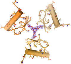
At the same time, Olga continued to contribute with her knowledge on photoregulation in aquatic photosynthesis (V. U. U. Chukhutsina et al., 2017; Van Den Berg et al., 2019). In early 2018, she moved to Imperial College (UK) to join the group in Jasper van Thor’s lab, first as an independent EMBO fellow and later as a Marie Curie fellow. This was her first project in crystallography.  She began to study a fascinating protein involved in the photoregulation of aquatic photosynthesis, called the orange carotenoid protein (OCP). Her stay at Imperial College led to the first light-induced OCP structure (Chukhutsina et al., 2022). OCP is a very difficult protein to study because it has an extremely low photoconversion rate, so it is difficult to get measurable amounts of the photoconverted protein.

Currently Olga focusses her research on the photocycle of the OCP and its potential applications in environmental engineering. By gaining control over the photocycle of OCP, she believes it is possible to regulate the aquatic biomass of microorganisms. This could have significant implications for ecological management, particularly in marine environments. Primarily her work centers on photosynthesis and photoregulation in cyanobacteria and brown algae, which are key contributors to aquatic biomass. These organisms thrive in extreme conditions, and Olga is investigating their behavior in vivo through advanced spectroscopic methods. Her goal is to understand how they manage light and stress, which in turn influences their growth and survival. Another exciting area of Olga’s research centers on special algae known as dinoflagellates, which live in symbiosis with corals. These algae play a crucial role in coral health, as corals rely on the high-energy metabolites, like sugars, produced by dinoflagellates. When these algae are exposed to stressors such as high temperatures, excessive light, or increased acidity, coral bleaching can occur, which poses a major threat to coral reefs and the biodiversity they support. As coral reefs account for 25% of oceanic biodiversity, understanding the relationship between algae and corals is critical to conservation efforts.

To delve deeper into the mechanics of OCP and its role in photoregulation, Olga combines advanced spectroscopy with structural biology. She frequently utilizes cutting-edge serial crystallography techniques at world-renowned facilities such as the Diamond Light Source, LCLS, MAX IV, and SwissFEL. These methods enable her to explore the structural dynamics of OCP in great detail, with the hope of uncovering mechanisms that could be leveraged to optimize photosynthesis. A unique aspect of Olga’s research is her synergetic approach, which always connects structural changes observed in the lab back to the larger biological context. She grows and studies photosynthetic organisms in her lab while also examining the behavior of isolated protein crystals, providing a comprehensive view of how these processes function in nature.

Looking ahead, Olga plans to continue using these advanced crystallographic approaches to study other key photosynthetic regulators that are activated by light or pH changes. Her ultimate aim is to understand how these proteins control photoregulation on a molecular level.

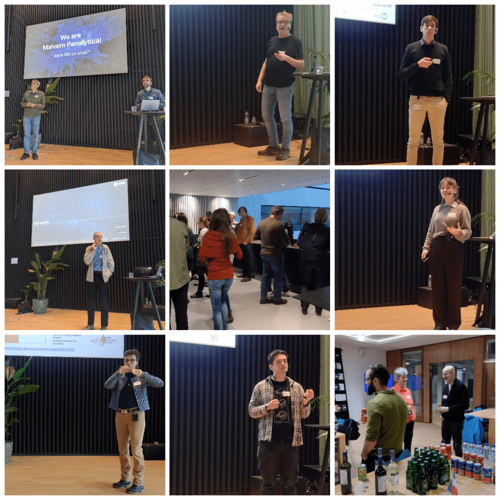
In 2023 Olga got Unesco Loreal Women in Science Prize and was a NIAS (Netherlands Institute for Advanced Science) fellow 2023-2024. Olga will organize a workshop in June 2025 on the ‘Effect of Temperature on Photosynthesis’ – NVK will report on this and let you know.

Currently there is a postdoc opening in her group, get in touch via v.u.chukhutsina@vu.nl


References:

Bag, P., Chukhutsina, V., Zhang, Z., Paul, S., Ivanov, A. G., Shutova, T., … Jansson, S. (2020). Direct energy transfer from photosystem II to photosystem I confers winter sustainability in Scots Pine. Nature Communications, 11(1), 1–13. https://doi.org/10.1038/s41467-020-20137-9

Chukhutsina, V. U. U., Fristedt, R., Morosinotto, T., & Croce, R. (2017). Photoprotection strategies of the alga Nannochloropsis gaditana. Biochim Biophys Acta, 1858(7), 544–552. https://doi.org/10.1016/j.bbabio.2017.05.003

Chukhutsina, V. U. V. U., Holzwarth, A. R. A. R., & Croce, R. (2019). Time-resolved fluorescence measurements on leaves: principles and recent developments. Photosynthesis Research, 140(3), 355–369. https://doi.org/10.1007/s11120-018-0607-8

Chukhutsina, V U, Baxter, J. M., Fadini, A., Morgan, R. M., Pope, M. A., Maghlaoui, K., … van Thor, J. J. (2022). Light activation of Orange Carotenoid Protein reveals bicycle-pedal single-bond isomerization. Nat Commun, 13, 6420. Retrieved from http://biorxiv.org/content/early/2022/09/05/2022.01.17.475858.abstract

Chukhutsina, Volha U., Liu, X., Xu, P., & Croce, R. (2020). Light-harvesting complex II is an antenna of photosystem I in dark-adapted plants. Nature Plants, 6(7), 860–868. https://doi.org/10.1038/s41477-020-0693-4

Van Den Berg, T. E., Chukhutsina, V. U., Van Amerongen, H., Croce, R., & Van Oort, B. (2019). Light acclimation of the colonial green alga botryococcus braunii strain showa. Plant Physiology, 179(3). https://doi.org/10.1104/pp.18.01499

Written by Sven Hennig

Save the dates for upcoming NVK events!

We hope you had a good summer season and wanted to briefly announce a few upcoming events for the second half of the year.

NVK Crystallography Course in Utrecht at 15.-17.01.2025

The NVK organizes a crystallography & structure determination course in January in Utrecht under the guidance of Loes Kroon. The course is suitable for newcomers in the field, they can choose between powder and single crystal diffraction. As soon as a program is available we will announce it on social media and our website. Please save the date for this, if you are interested in sending some of your PhD students, Postdocs or research assistances.

Algemene Leden vergadering (ALV) 2024 on the 25.11.2024 in an online fashion

We will inform you via email and website about the detailed program. With the final invitation we will circulate the minutes, program and the financial report. Also you will find a zoom link for the meeting. If you want to test your camera, microphone and connection in a dry run, I will provide a test link on Friday 22.11.2024, 11:00-11:30. You can log in, check & leave, there will be no content in this session.

Please feel free to save the date for those two events and we are looking forward to seeing you there.

Registrations for our spring symposium are open!

We are pleased to join forces with the Dutch Association for Crystal Growth (DACG) for the Spring Symposium. The event will be hosted by Maastricht University on May 30 & 31, 2024, and registrations are open till May 15, 2024.

This event aims to provide ample opportunity for fruitful discussion and dialogues between experts from different fields of crystallization and to incite collaborations at the international level between the different disciplines on the area of nucleation, crystallization, crystal growth and self-assembly.

An early career event will be hosted on May 30, focusing on MSc, PhD students and postdocs future career opportunities. All participants to the early career gathering are kindly invited to bring a poster about their research. Please send your abstract latest on May 15th, 2024 at info@dacg.nl. We will have two poster sessions dedicated to networking and brainstorming. Please indicate in your registration if you will be taking up this opportunity. The posters will remain in display throughout May 31st.

The DACG & NVK Spring Symposium will take place on May 31 featuring invited talks by renown researchers from the Netherlands and neighboring countries, contributed talks.

We hope to welcome you, please register here.

Sven Hennig and colleagues published on covalent bicyclization of quaternary protein complexes

Congratulations to our member Sven Hennig and his colleagues Tom Grossmann (Biomimetic Chemistry) from the Vrije Universiteit Amsterdam and Saskia Neubacher (incircular) for the publications on the covalent bicyclization of quaternary protein complexes in Chem. Great work in which they show not only the successful crosslinking and stabilization of the Pseudomonas fluorescens esterase (Pfe) trimer via the INCYPRO technology, but also solved its crystal structure (PDB ID 8pi1).

Save the date! – Joint meeting NVK and DACG

A joint symposium of NVK together with DACG (Dutch Association for Crystal Growth) will be organized at the Science Campus of Maastricht University on 31 May 2024. On the afternoon before, 30 May,  we will organize a satellite meeting for young researchers with an interesting evening program and overnight stay.  Details following soon.

Merry Christmas and a happy New Year!

We wish you a very pleasant and peaceful Christmas and a Happy New Year. We hope for an interactive, inspiring and fruitful 2024!

We hope to see you at our spring-symposium, stay tuned for updates!

NVK symposium was a great success!

On 24.11.2023 we held our annual general meeting of the NVK (ALV) at the Amsterdam Science Park organized by Roland Bliem and the ARCNL institute. Apart from the official ALV, the day was full of scientific talks and exchange, as we embedded it into our autumn symposium, this year on the topic of ‘Crystallography in Materials’. We had an exciting panel of speakers especially Ruud den Adel (Unilever), Tom de Vries (Nijmegen), Mirijam Zobel (Aachen), Alexey Pustovarenko (Malvern Panalytical), Adrian Graham (ESA/ESTEC) and Jarek Mazurek (Ardena).

A sphere impression of the day!

Special thanks belong to our speakers of the young researcher session (Grisha Shipunov (UvA), Falk Pabst (UvA) and Charlotte Vrolijk (Maastricht)), which was a great success and gave our next generation researcher a platform to discuss their achievements.

IMG_20231124_120220794_HDR

Image 1 of 27

Find the full program here. If you got curious, please join our next Symposium in Spring 2024! Details will follow.